
If your doctor finds a uterine cyst on your ultrasound, you’re likely asking, “Now what?” It’s natural to feel uneasy when something unexpected shows up on a scan. While many cysts are harmless, knowing what the imaging shows in uterine cyst ultrasound findings can help you feel more in control. We’ll cover what those findings mean, what changes may need attention, and how your doctor might respond.
Key Takeaways:
- A uterine cyst is a fluid-filled area in the muscle wall of the uterus; however, they are common and often harmless.
- Causes can include hormonal shifts, past injuries, endometriosis, or fibroid breakdown. Consequently, many women experience these conditions.
- They are diagnosed mainly through ultrasound: transvaginal (more detailed) or transabdominal (wider view), providing a comprehensive examination.
- Simple cysts look black and have thin walls; in contrast, complex ones might show septations, echoes, or blood flow.
- Importantly, functional cysts often form during the menstrual cycle and usually disappear on their own.
- Doppler ultrasound checks blood flow; increased flow may be a concern.
- Cysts larger than 4 cm, with complex features or fast growth, need closer review or treatment. Therefore, monitoring is essential.
- Distinct features help separate cysts (fluid-filled) from fibroids (solid) or polyps (having a central blood supply). This distinction guides treatment.
- Symptoms may include pelvic pain and irregular or heavy periods. As a result, these symptoms should be assessed by a doctor.
- Follow-up is critical—suspicious findings may lead to biopsy or surgery.

Understanding Uterine Cyst Ultrasound Findings and What They Mean for You
Uterine cysts are more common than many people realize. Often discovered during routine exams or when investigating symptoms like pelvic pain or irregular bleeding, these fluid-filled sacs can appear inside the muscle of the uterus. While most are harmless and go unnoticed, uterine cyst ultrasound findings provide key insight into your reproductive health and guide what happens next.
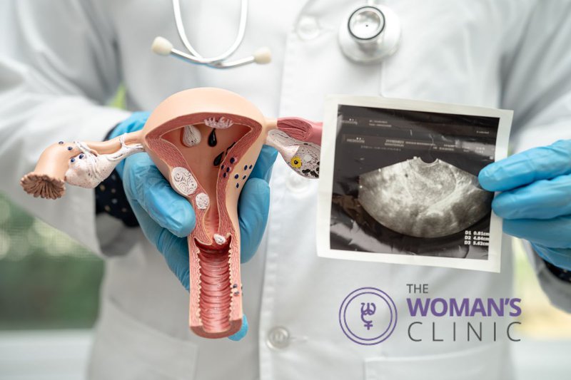
At The Woman’s Clinic in Little Rock, we use advanced ultrasound imaging to identify, monitor, and explain uterine cysts so you feel informed and supported at every step.
How Uterine Cysts Are Detected and Diagnosed
A uterine cyst is a small fluid pocket that develops in the muscular layer of the uterus. These cysts are usually found during scans ordered for symptoms such as pelvic pressure, pain, or abnormal bleeding. Many people with these cysts don’t even know they have them until imaging picks them up.
To detect cysts, your provider will likely start with ultrasound imaging—a safe and noninvasive method that uses sound waves to visualize the uterus and surrounding organs. Typically, there are two main types of ultrasounds used:
- Transabdominal ultrasound, performed through your lower belly
- Transvaginal ultrasound, done using a probe inserted into the vagina for a closer, clearer view
Transvaginal ultrasound is especially useful for identifying small or subtle changes within the uterine wall. On the screen, a cyst typically appears as a round, dark (anechoic) area; this darkness indicates fluid. Furthermore, thin, smooth walls and clear contents usually point to a benign finding.
What Causes Uterine Cysts to Form
Uterine cysts can form for several reasons, including:
- Hormonal changes, such as those seen during your menstrual cycle or menopause
- Scarring from surgery or childbirth
- Endometriosis, where uterine-like tissue grows outside the uterus
- Degeneration of fibroids, when fibroids break down and form cystic spaces
Not all cysts require treatment. But when they become painful, bleed internally, or show complex features on imaging, further evaluation is needed.
How Types of Uterine Cyst Findings Appear on Ultrasound
When reviewing uterine cyst ultrasounds, providers look for several key features to determine the type and risk level of a cyst:
- Shape and borders: A simple cyst is usually round with smooth edges
- Echogenicity: This describes how much sound the cyst reflects
- Internal contents: Clear fluid is a good sign; solid tissue or debris may require more attention
Simple vs. Complex Cysts
- Simple cysts appear dark, thin-walled, and filled with fluid
- Complex cysts may include thick walls, septations (internal divisions), or echogenic material (suggesting blood or tissue)
Complex cysts are not always dangerous, but they do prompt closer observation or follow-up. Read more on Radiopaedia’s imaging guide for visuals.
Functional Cysts
Some cysts are functional; importantly, they form as part of your normal cycle. Typically, these tend to be small, clear, and temporary. Consequently, they often resolve on their own without requiring treatment.
Others, especially those linked to endometriosis or fibroid degeneration, appear more persistent and may cause ongoing symptoms like pain or abnormal bleeding.
Why Follow-Up Imaging Matters
If a cyst is found on your ultrasound, your doctor may ask for follow-up imaging in 6 to 8 weeks. This repeat scan is important as it helps determine whether the cyst has shrunk, remained the same, grown larger, or changed in appearance. Consequently, stable or shrinking cysts are typically left alone. Meanwhile, growing or complex cysts may require additional imaging or tests to rule out rare causes, such as malignancy.
- Has shrunk
- Stayed the same
- Grown larger
- Changed in appearance
Stable or shrinking cysts are typically left alone. Growing or complex cysts may require additional imaging (like MRI) or lab tests to rule out rare causes such as malignancy.
Key Ultrasound Findings That Help Identify a Uterine Cyst
One of the main reasons we rely on uterine cyst ultrasound features is to effectively distinguish between various types of uterine abnormalities. Transition words like "for instance," "therefore," and "in addition" can be used throughout the process to guide and clarify the differentiation needed for accurate diagnosis and treatment.
Echogenicity
- Low echogenicity (dark appearance) = clear fluid inside
- High echogenicity (bright appearance) = thicker fluid, blood, or tissue
- Mixed echogenicity = suspicious or complex cysts needing close review
Internal Echoes and Septations
- No echoes = likely benign
- Internal echoes = possible debris or clotted blood
- Septations = internal walls may indicate complexity
Doppler Flow Patterns
Using Doppler ultrasound, we can see if blood is flowing in or around the cyst:
- No blood flow = likely inactive, benign cyst
- Active flow = may indicate growth or activity, which requires attention
Learn more about how these features help direct diagnosis in this ultrasound review of uterine lesions.
Comparing Transvaginal and Abdominal Ultrasound
Transvaginal ultrasound is often more accurate for detecting uterine cysts, especially small or internal ones. It provides:
- Better resolution
- Clearer view of the uterine lining and muscle
- More accurate measurement of cyst size and structure
Meanwhile, abdominal ultrasound is helpful for viewing larger cysts or those located outside the uterus. In some cases, both types are used together for a full view.
Distinguishing Cysts from Other Uterine Conditions
On ultrasound, cysts must be differentiated from:
- Fibroids – solid, dense tissue masses (appear gray or mixed)
- Polyps – small growths within the lining, usually with one blood vessel
- Ovarian cysts – may look similar but are located on the ovary, not in the uterus
Movement during the scan, location, and how the cyst interacts with the uterine wall help determine where it originates. When in doubt, your provider may order additional imaging or a specialist review.
Common Symptoms Linked to Uterine Cysts
While many uterine cysts cause no symptoms, you might experience:
- Pelvic pain or pressure
- Heavy or irregular bleeding
- Bloating or fullness
- Cramping that doesn’t go away
Changes in your menstrual cycle—like skipped periods, spotting, or unusually long flow—can also occur if the cyst disrupts hormonal signals.
If pain is persistent or severe, or if bleeding becomes heavy, ultrasound can help clarify the cause.
When Uterine Cyst Ultrasound Findings Suggest Concern
Certain ultrasound markers may prompt a closer look. These include:
- Thickened cyst walls
- Solid components inside
- Irregular blood flow on Doppler
- Rapid growth or size over 4 cm
These signs don’t always mean cancer, but they do require further evaluation. In some cases, if the findings raise concerns, a biopsy or referral to a gynecologic oncologist may be recommended, especially for postmenopausal patients, since their hormone levels change the way cysts behave.
What to Expect from Follow-Up and Treatment
If your cyst appears simple and you’re not having symptoms, watchful waiting is often the first step. Therefore, this means repeating the scan in a few weeks and monitoring for any changes. This approach ensures that you remain informed about your condition. Moreover, it helps your doctor decide if any further action is necessary.
Follow-Up May Include:
- Repeat ultrasound
- Blood tests (like CA-125) if malignancy is a concern
- Doppler scan to reassess blood flow
- Discussion of symptoms and medical history
When Treatment Is Needed
Treatment may be necessary if the cyst:
- Continues to grow
- Causes ongoing pain or pressure
- Has complex features
- Appears after menopause
- Interferes with fertility or menstrual cycles
Treatment options include:
- Hormonal therapies to reduce cyst formation
- Surgical removal of the cyst or affected area
- Myomectomy (if the cyst is part of a degenerating fibroid)
Your care plan is always based on your symptoms, goals, and the overall ultrasound findings.
Interpreting Your Pelvic Ultrasound Report
Ultrasound reports often include terms like:
- Anechoic: dark, fluid-filled, likely benign
- Hypoechoic: less dark, may contain blood or tissue
- Septated: divided sections inside
- Echogenic material: solid or complex components
Discuss these findings with your provider during your visit. Ask how they match your symptoms and what the safest next step is for you.
Follow the Next Step Toward Clarity and Care
If you’ve received ultrasound results or are experiencing symptoms like pelvic pain or irregular bleeding, we’re here to help. At The Woman’s Clinic in Little Rock, our team offers expert imaging, clear explanations, and personalized care plans that fit your needs. Whether you’re monitoring a simple cyst or exploring treatment options, you’ll receive compassionate, evidence-based support every step of the way. Schedule your appointment today to get the answers and care you deserve.